
Click the image above to view full, printable PDF of this article.
By Abby Flyer, M.A., Communications and Education Specialist. Originally published in LEMURS Magazine: Reasons for Hope in February 2026.
When caring for the world’s most endangered group of mammals, no cure is “one size fits all.”
Between 55 and 25 million years ago, the ancestor of aye-ayes and the ancestor of all other lemurs were blown out to sea, drifting across the Mozambique channel from mainland Africa and landing on the island of Madagascar.
For the next tens of millions of years, in a process called adaptive radiation, these primates diversified and spread across the island, developing different strategies for survival based on geographic region, available resources, and competition. Today, lemurs are a remarkably diverse group of primates. Currently there are more than 100 different species of lemur, in addition to at least 17 species that have already gone extinct.
The Duke Lemur Center houses the most diverse population of lemurs on Earth outside their native Madagascar. Not only are we committed to meeting the needs of the 10 lemur species under our care, every individual lemur—nearly 250 of them!—receives an individualized diet plan, enrichment, and veterinary care. And just like humans, no two lemurs are exactly alike.
From neonatal care to accommodations for our oldest residents, these six individuals highlight the extraordinary measures that the DLC’s animal care team takes to ensure that every single animal in our colony receives the highest quality of care.
Cirilla
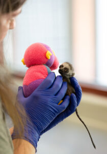
Cirilla at seven days old during a neonatal exam. Photo by Sara Sorraia.
Ring-tailed lemur Cirilla was born weighing just 37 grams and, at barely half the weight of an average 70-gram infant, is tied for the second smallest surviving ring-tailed lemur birth in DLC history. Her temperature was low and she had difficulty holding up her head and clinging to mom Alena. Mom and baby were rushed to the Anna Borruel Codina Center for Lemur Medicine and Research and into the ICU incubator.
Veterinary and husbandry staff took turns positioning Cirilla to nurse on Alena every two hours, throughout the day and night. Cirilla also received oral and subcutaneous doses of dextrose and was held against a heat pack to help her maintain an appropriate internal body temperature.
After three full days in the ICU, Alena and her daughter returned to their home enclosure, where they remained under close supervision. Cirilla had gained four grams and was able to nurse without intervention. By four days old, Cirilla’s eyes finally opened.
Over a year later, Cirilla has grown into a thriving adult lemur. While she remains small in stature, Cirilla has a big personality and is eager to explore the forest as she free-ranges with her mom, dad, and older brothers.

Cirilla free-ranging this summer. Photo by Sara Nicholson.
Ozma
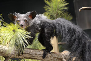
Photo by David Haring.
When Ozma, a 34-year-old aye-aye, developed a dental abscess on the right side of her jaw, the DLC veterinary team drained and treated it, as usual, with antibiotics. When the infection didn’t clear, she was driven to the Veterinary Dental Clinic of North Carolina, where the clinic’s owner, Don Hoover, D.V.M, had offered the use of his small animal CT scanner. Dr. Hoover’s CT scans allowed the team to visualize Ozma’s problem more clearly: She had a major tooth fragment on the right side of her mouth.
After determining that a 3-D print would allow better visualization of the problem and more precise surgical planning, the team met with Chip Bobbert, a Digital Fabrication Architect at the Duke University Office of Information Technology. Bobbert’s team agreed to create the model in their 3-D printing lab on campus and soon delivered a prototype to the vet lab, but printing in a single color made it difficult for the veterinary team to see what was tooth and what was bone. So the DLC veterinary team worked with Susan Whitney, who converts CT scans into 3-D printable files, to create an improved two-color model of Ozma’s mandible.
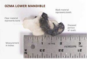
3-D printed lower mandible of Ozma. The DLC vet team collaborated with Chip Bobbert and Susan Whitney, who donated their time to convert Ozma’s CT scans into 3-D printable files. Because the resulting 3-D model was true-to-size and -form, the veterinarians could better visualize Ozma’s problem and plan her surgery more precisely. Photo by Sara Sorraia.
The two-color model helped the vets better identify what was tooth, what was abscess, and what was bone. The model was so detailed and true-to-life, it even enabled the vets to determine where to administer lidocaine before making the extraction.
Ozma’s heart rate did not increase at all during surgery, indicating that the nerve block had worked and she was feeling no pain. The entire surgery—from placing the anesthesia monitors and the IV catheter, to administering the nerve block, extracting the tooth, instilling the antibiotic gel, suturing the surgery site, and turning off the anesthesia—took only an hour.
Aristides
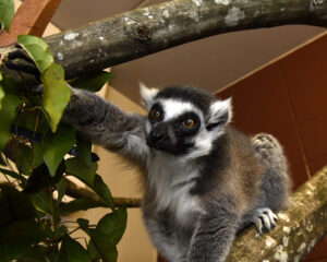
Photo by David Haring.
When it comes to branching geriatric lemurs’ enclosures, the DLC’s husbandry technicians need to take extra precautions. Ring-tailed lemur Aristides lived to 32 years old and spent the latter years of his life with degenerative joints, making it more difficult for him to jump and move about his enclosure. Sarah Midolo, one of Aristides’ primary caretakers at the end of his life, always considered Aristides’s physical limitations when building his structural enrichment.
“I gave him a lot of textured, thick branches so he had something to grab onto,” she explains. “I made sure that he had an avenue everywhere and that he didn’t have to jump for any of it, that he could just walk along the branches kind of like a big ramp system.”
The doors between Aristides’s indoor and outdoor enclosures were too high, so she installed mesh and metal ramps to help him transition smoothly between the two environments. Sarah used twice as many zip-ties to secure each of his structures. “I made sure everything was extra supported. I didn’t want anything wobbling and him falling.”
Kizzy
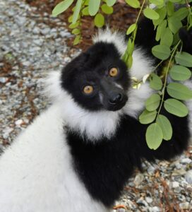
Photo by David Haring.
20-year-old Kizzy, a black-and-white ruffed lemur, has developed arthritis in her hips and elbow. In addition to a joint supplement, Kizzy has begun photobiomodulation therapy with the DLC’s veterinary technicians. This laser therapy is a non-invasive treatment that can help decrease inflammation and swelling, speed up healing, and encourage blood flow to the area treated. To the lemur, it just feels like a gentle touch with the warm laser head, and Kizzy participates voluntarily during training sessions.
“Each species of lemur has different qualities that you have to account for in treatments,” explains Cat Ostrowski, R.V.T., one of the DLC’s vet techs. “Skin color, size, ability to be involved in training… One species may love to be on the ground, so we will train on the ground; but not all species are comfortable at ground level, so we use their natural history to inform us of what positions we can ask for during a training session and then build our treatment sessions around that.” Currently, Kizzy receives laser therapy on her hips twice a week.
Motmot
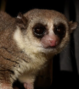
Photo by David Haring.
At 12 years old, fat-tailed dwarf lemur Motmot was anesthetized for a veterinary exam when DLC staff noticed that her left nictitating membrane—a third eyelid present in many animals, including lemurs—was thicker and more prominent than normal. The vet team carefully collected a tissue sample, and upon inspection by a pathologist, the suspected diagnosis was cancer (later confirmed to be lymphoma).
To determine whether the eyelid required surgical removal, the DLC’s veterinary team consulted with Sophie Rajotte, D.V.M., M.S., DIPL. ACVO., a veterinary ophthalmologist at Animal Eye Care of Durham. The situation was uniquely complicated due to the time of year: Motmot was about to enter torpor. “Animals’ slowed metabolic rate during the torpor season means that healing can be slowed during this time,” explains DLC veterinarian Julie Ter Beest, M.S., D.V.M., DIPL. ACZM. The team elected to monitor the eyelid rather than attempt surgery and prescribed steroid drops to reduce inflammation.
After a couple of months, it became evident that surgery would be necessary. By this point, the torpor season was ending, and Dr. Rajotte was able to surgically excise the nictitating membrane with no complications. Two years later, Motmot still has routine checks to monitor her recovery, but so far, she has remained cancer free.
Virginia Creeper
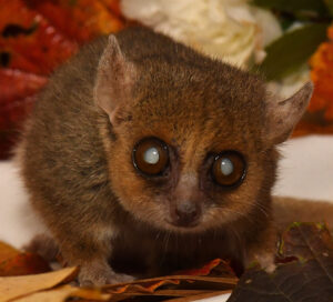
Photo by David Haring.
In their native Madagascar, gray mouse lemurs only live three to five years. Here at the DLC, these tiny primates can live into their early teens!
13-year-old Virginia Creeper is the oldest gray mouse lemur in the DLC’s colony. At this late stage in her life, Virginia Creeper struggles with decreased mobility, vision loss, and cognitive decline. Encouraging her to eat has been particularly challenging. “She’s been experiencing mandibular swelling,” explains Allie Monahan, one of the DLC’s husbandry technicians and the geriatric mouse lemur’s primary caretaker. “I soak her chow before feeding her to make it easier to eat.”
The idea came to Allie while planning a special birthday treat for Virginia Creeper, offering her gruel (primate chow mixed with water and a tasty flavoring, like honey or coconut milk) rather than her standard solid food. “When I gave it to her, she chowed down like I’ve never seen before, and I’ve been soaking her chow ever since! Since then, she’s been more consistent in her eating and has gained weight.”
Allie has to consider not just what she feeds Virginia Creeper, but how she feeds her. “I always feed her in the same place,” says Allie, which helps ensure that the lemur can consistently find her food bowl as her vision and memory decline. “Also, she’s only fed in flat dishes to make the food more easily accessible.”
Specialized Guts
Lemurs aren’t just diverse in appearance—their guts also vary in length and complexity. Longer relative digestive tracts allow a greater relative surface area for nutrient absorption. As a result, animals with a higher intestine length to body length ratio are able to digest more complex foods.
Coquerel’s sifakas (Propithecus coquereli), who are specialized leaf eaters, have a digestive tract nearly 16 times the length of their bodies. For comparison, the fruit-eating red ruffed lemurs (Varecia rubra) have guts approximately five times their body length.
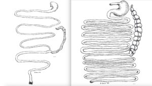
Gastrointestinal tract from a red ruffed lemur (left) and a Coquerel’s sifaka (right). Scale equals 1 cm. Diagrams reproduced from Campbell et al. 2000. “Description of the gastrointestinal tract of five lemur species: Propithecus tattersalli, Propithecus verreauxi coquereli, Varecia variegata, Hapalemur griseus, and Lemur catta.” American Journal of Primatology. 52: 133-142.
While a longer gut allows sifakas to digest complex plant matter efficiently, it also means that consuming excessively sugary fruit can upset the sifaka’s delicate gut system, making them extremely (and sometimes fatally) sick. Fruits from the forests of Madagascar, which make up a regular part of wild sifakas’ diets, have the approximate sugar content of an American-grown cucumber; whereas in the United States, many fruits and veggies are cultivated to maximize their sugar content. As a result, for the DLC’s sifakas, fruit is off the menu entirely. This is why it’s crucial to individualize diet plans—a healthy snack for a ruffed lemur could send a sifaka straight to the vet!
A longer gut also means more time to digest. Food can take up to 36 hours to pass through a Coquerel’s sifaka, while red ruffed lemurs go from eating to excreting in under two hours. Beyond allowing ruffed lemurs to enjoy more sugary produce, this allows whole seeds to pass through their digestive systems. Everywhere red ruffed lemurs travel, they drop seeds in their own little piles of fertilizer (aka poop), helping to disperse seeds for a variety of plant species across Madagascar’s tropical rainforests.
Photo by Sara Nicholson.